Digital X-Ray Imaging
What are X-rays?
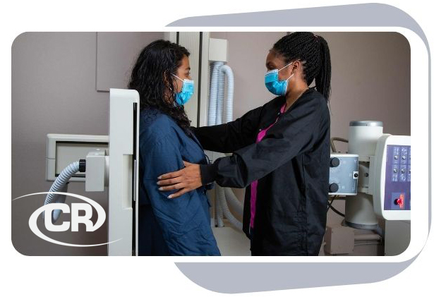
X-rays are a form of electromagnetic radiation, just like visible light. X-ray machines send individual x-ray particles, called photons. These particles pass through the body. At Clermont Radiology a computer is used to record the digital images that are created. X-ray imaging is still the most commonly used technique in radiology.
To make a radiograph, a part of the body is exposed to a small quantity of x-rays.
X-rays are safe when properly used by a radiologist and technologist specially trained to minimize exposure. No radiation remains afterward. X-rays are used to image every part of the body.
An X-ray machine is essentially a camera. Instead of visible light, however, it uses X-rays to expose the image. X-rays are like light in that they are electromagnetic waves, but they are more energetic so they can penetrate many materials to varying degrees. Since bone, fat, muscle, tumors and other masses all absorb X-rays at different levels; you see different shaded structures on the image.
How should I prepare?
For 6 hours prior to the procedure do not eat food, suck on candy, or chew gum. Drink plenty of water; it keeps your vascular system hydrated. Limit your physical activity 24 hours before the exam. To ensure the best results are acquired do not wear clothing with zippers, snaps, or metal attached. If insulin dependent, take ½ 4 hours prior to exam and bring the other ½ to take after the procedure. If oral diabetic medication is needed, don’t take the morning dose; bring it with you to take after the procedure.
What should I expect?
The technologist will explain the procedure to you, check your glucose levels, and answer any question you may have. Once injected with the radiotracer you will sit in a recliner with blankets to allow the dose to uptake in your system for 60 minutes. You will then be imaged for 20 – 40 minutes depending on the specifications of the exam your doctor requested.
How do I get my results?
After you study is complete, our board certified radiologists will evaluate the images from your PET scan and send a complete report to your doctor, who will discuss the results with you.

Clermont News
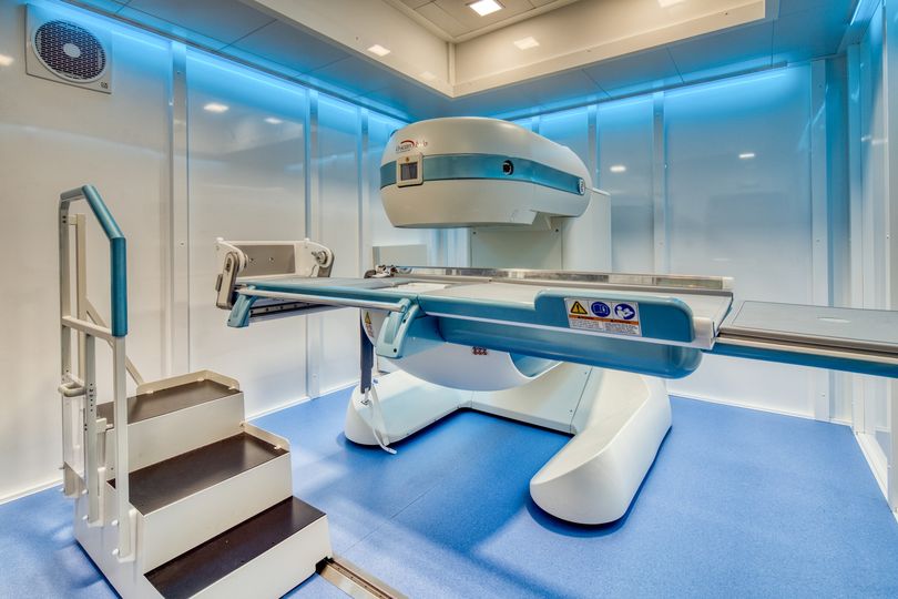
Our Esaote G-Scan Brio MRI located in our Ocala office!
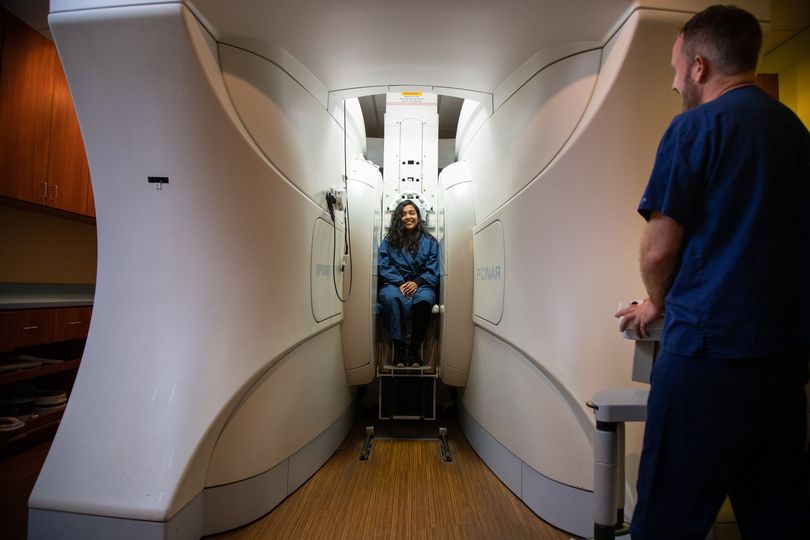
Oakley Seaver Open MRI
The 3 “Must Haves” in Women’s Imaging
Make an Appointment
Filling out the form does not guarantee an appointment until confirmed via phone or email by a patient care representative.
In a continued effort to improve patient care, we will now require all orders on file prior to scheduling for the following exams:
- MRI
- CT
- PET
- Nuclear Medicine
Clinical notes are needed prior to requesting authorization. Any delay in receiving the necessary notes may result in the rescheduling of appointments.


