Short Bore High Field MRI (Magnetic Resonance Imaging)
Clermont Radiology is a Magnetic Resonance Imaging Accredited Facility by the American College of Radiology (ACR).

What is Magnetic Resonance Imaging?
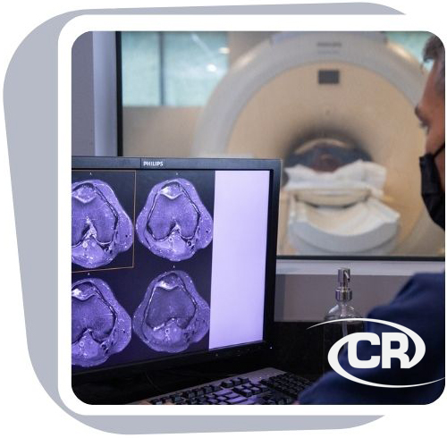
Magnetic Resonance Imaging, MR or MRI is an advanced imaging method that produces images of the body without surgery or x-rays. Magnetic Resonance Imaging utilizes the forces of magnetism and radio waves to create images or pictures of the human body.
You are placed in a strong magnetic field, which causes the hydrogen atoms in your body to align in a positive position to receive radio signals. Your body then transmits signals of its own which are collected and processed by a computer then transformed into the images that our radiologist will read.
Available at these locations:

Advanced Technology. Every MRI scanner has what is called “field strength.” Field strength is the power of the scanner’s magnet. With higher field strengths, your scan pictures are clearer and show smaller details of your body. Clermont Radiology uses a High Field MRI.

Sometimes referred to as a "Closed MRI," these types of units consist of a much stronger magnetic field, which provides for better overall image quality and increased speed of the examination. Due to the cylindrical configuration of the unit, the patient is fully surrounded by the magnetic field, which allows us to provide high-resolution images in a fraction of the time.
Tell the technologist if any of the following pertain to you:
-
You have a pacemaker
-
History of working with metal
-
Brain aneurysm clips
-
History of injury during Military service
-
Metallic plates or other implants
-
Nursing or chance you could be pregnant
What should I expect?
The MRIexam can be one of the easiest and most comfortable exams to experience. The technologist will ask you to lie on a cushioned table and often an imaging device called a "coil" will be placed around the area of the body to be scanned. Once you are comfortably positioned, the table will move into the magnet opening. As images are acquired, you will hear “knocking” or “buzzing” sounds for a few minutes at a time.
It is important to lie as still as possible during this part of the exam to help us capture clear images. If necessary, physician-administered medication is available to help you relax. In some cases, you will need contrast material to further aid in detection or diagnosis of potential abnormalities.
How should I prepare?
Usually, there is no preparation prior to a MRI exam. Any metal and jewelry such as earrings, glasses, or hairpins should be removed.
How do I get my results?
After your study is complete, our board certified radiologist will evaluate the image results and send a full report to your doctor, who will discuss the results with you.
Clermont News
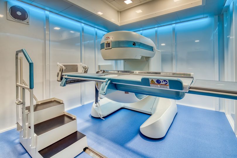
Our Esaote G-Scan Brio MRI located in our Ocala office!
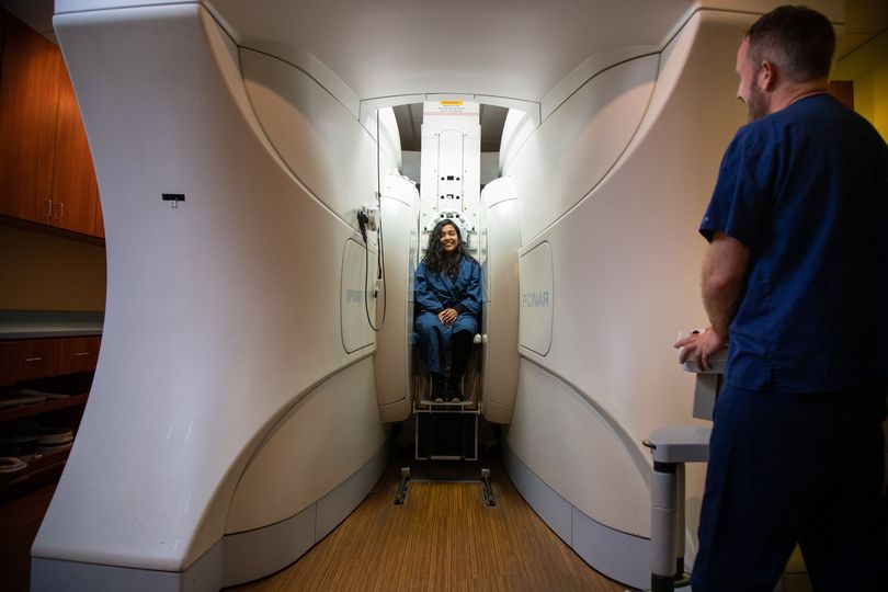
Oakley Seaver Open MRI
The 3 “Must Haves” in Women’s Imaging
Make an Appointment
Filling out the form does not guarantee an appointment until confirmed via phone or email by a patient care representative.
In a continued effort to improve patient care, we will now require all orders on file prior to scheduling for the following exams:
- MRI
- CT
- PET
- Nuclear Medicine
Clinical notes are needed prior to requesting authorization. Any delay in receiving the necessary notes may result in the rescheduling of appointments.


