Stand Up MRI (Magnetic Resonance Imaging)
Clermont Radiology is a Magnetic Resonance Imaging Accredited Facility by the American College of Radiology (ACR).

What is Magnetic Resonance Imaging?
Magnetic Resonance Imaging, MR or MRI is an advanced imaging method that produces images of the body without surgery or x-rays. Magnetic Resonance Imaging utilizes the forces of magnetism and radio waves to create images or pictures of the human body.
You are placed in a strong magnetic field, which causes the hydrogen atoms in your body to align in a positive position to receive radio signals. Your body then transmits signals of its own which are collected and processed by a computer then transformed into the images that our radiologist will read.
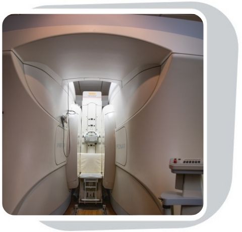
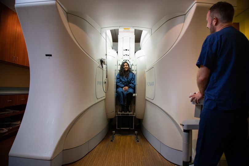
For patient who are claustrophobic (have extreme difficulty and are wary of being confined in small spaces), the Upright MRI is the perfect solution. The Upright MRI allows imaging of the spine in an upright, weight-bearing position and views the curvature of the vertebrae and soft tissues including the nerves and discs.
The Upright MRI has the ability to put the patient in the position necessary to give the most complete and accurate diagnosis. As a result of its unique ability to scan patients in weight-bearing positions, the Upright MRI has detected problems that would have gone undetected on ordinary lie-down scanners.
Tell the technologist if any of the following pertain to you:
-
You have a pacemaker
-
History of working with metal
-
Brain aneurysm clips
-
History of injury during Military service
-
Metallic plates or other implants
-
Nursing or chance you could be pregnant

What should I expect?
Depending on the body part being evaluated and the pain you are experiencing, you can be scanned in a multitude of positions standing, sitting, lying down or bending. Our technologist will help position you in the scanner. An imaging device called a "coil" will be placed around the area of the body to be scanned.
Please Note: Our Upright unit can accommodate patients up to 450 pounds, but height plays a role in patient fitting. A fitting may be needed if the patient’s weight is 300-350 pounds and their height is less than 5’5".
In some cases, you will need contrast material to further aid in detection or diagnosis of potential abnormalities. In this instance, an I.V. will be placed in your hand or arm. Once you are comfortably positioned, the chair will move into the magnet opening.
As images are acquired, you will hear a series of "knocking and buzzing noises" for a few minutes at a time. It is important to remain as still as possible during this part of the exam to help us capture clear images. Our technologist will be able to see and hear you. They will talk you through every stage of the exam.
How do I get my results?
After your study is complete, our board certified radiologist will evaluate the image results and send a full report to your doctor, who will discuss the results with you.
Clermont Radiology is a Center of Excellence for Imaging of the Spine.
Clermont News
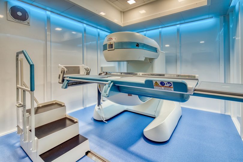
Our Esaote G-Scan Brio MRI located in our Ocala office!
Oakley Seaver Open MRI
The 3 “Must Haves” in Women’s Imaging
Make an Appointment
Filling out the form does not guarantee an appointment until confirmed via phone or email by a patient care representative.
In a continued effort to improve patient care, we will now require all orders on file prior to scheduling for the following exams:
- MRI
- CT
- PET
- Nuclear Medicine
Clinical notes are needed prior to requesting authorization. Any delay in receiving the necessary notes may result in the rescheduling of appointments.


